いよいよ関東も梅雨入りしたそうですね。
梅雨入りといえば、ほねゆきは師匠のあの言葉を思い出します。
師匠「ふつうの接骨院は梅雨は客足が遠のくが、骨折をしっかり診ている院は逆に患者さんが多くなる。」
おそらく、雨になると滑って転倒したり、遠くの病院へ行くより接骨院が近くて雨の中でも行きやすい(昔は今より病院数が少なかった)という理由で師匠は言っていたと思います。
現代では、この言葉がそのまま当てはまるかどうかは分かりませんが、やはりなぜかほねゆきの接骨院は梅雨になると患者さんが増えます。
ほねゆきは、今はあまり自費だけで患者さんをみることはしない(療養費算定できる外傷しかみない)ので骨折患者さんばかりですが、ほねゆきの院においては師匠の言葉は当てはまっています。
みなさんの院はどうでしょうか?この機会に統計を取りたいです!
もし、「私の院も患者さん増えます!」という方がいれば、このブログ記事をSNSでシェアしてください!
逆に「いやぁ、患者さん減るなぁ」という方は、このブログ記事をSNSでシェアしてください!
そうすると、ほねゆきが喜びます!(笑)
さて、前々回のブログではColles骨折は「なんか、得体の知れない物だよ」みたいな話をしましたが、今回はいよいよ本物のCollesさんの文献が登場です。
この記事ではこれ以降、コーレスさんの物語が始まりますので、しっかりと注意書きを読んだ上で読み進めてください。
この記事はコーレス骨折について医療関係者向けに書いた非常にマニアックな記事となっております。骨折マニアだけこの先を読んでください。「マニアックすぎるよ!時間返せ!」というクレームは一切受け付けませんので、ご注意ください。笑
目次
- Abraham Colles(エイブラハム・コーレス)
- Edinburgh Medical and Surgical Journal(エジンバラ外科医学誌)
- 『On the fracture of the Carpal extremity of the Radius(手関節付近の骨折について)』
- ほねゆきの要約
- ほねゆきの感想
- コーレスさんが発表した骨折とは?
- まとめ
もくじ
Abraham Colles(エイブラハム・コーレス)
Abraham Colles(エイブラハム・コーレス)さんは1773年7月23日にイギリスのアイルランドのとてもお金持ちの家に生まれました。
当時に医学を勉強できる人というのは、ほぼお金持ちの家に生まれています。クールで器用な性格だったそうです。
ある日、コーレスさんの実家の近くのお医者さんの家が洪水で流されてしまい、その家の片付けを手伝っている時に解剖学の本を見つけます。
これが、コーレスさんと医学の出会いでした。
1790年(当時18歳)でコーレスさんはダブリン大学に入学し、いろいろあったのちにエジンバラ医科大学に行き、1795年に医学の修士をとります。
その後、ロンドンの外科医のところに修行に赴(おもむ)き、鼠蹊部の解剖に取り組むなどして、さらに医学の見地を広げていきます。
イギリスでしっかりと医師法が成立したのが1858年ですから、それよりも前に活躍されていた、医師のような立場の方だったんですね。
ちなみに、X線が発見(報告)されたのが1895年ですから、それよりも前に生きた先生がコーレス骨折について発表したとすると、これは当時からすれば非常に先進的だったのでしょう。(←後でまた出てくる話題だから覚えといてね!)
1804年から30年以上も外科医・大学教授として活躍し、1843年11月16日に没します。死因は痛風だったと記録がなされているようです。
また、今回は脱線しすぎな話題になるのであまり深くは書きませんが、鼠径靭帯もこのコーレスさんが発見したとか、鎖骨下動脈の結紮に世界で初めて成功したとか、会陰部の筋層についてはっけんしたりとか、いろいろと仕事してます。(笑)
Edinburgh Medical and Surgical Journal(エジンバラ外科医学誌)
Edinburgh Medical and Surgical Journal(エジンバラ外科医学誌)というのは、Collesさんがコーレス骨折を発表した雑誌の名前です。
この雑誌は1805年〜1855年に発刊されており、イギリスのスコットランドでは非常に有名な医療系雑誌でした。
イギリスでは1800年ごろに印刷機が発明されたといいますから、雑誌等の発刊の全盛期だったと言え、医学を学ぶ人らにとっては書物を読む習慣はある程度根付いてきていたようです。
1800年代というと、日本では出島にシーボルト(ドイツの医師)がシーボルト事件を起こしたり、歌川広重が東海道五十三次を完成させたり、開国したりとか騒がしい時代です。
そんな時代にCollesさんはいそいそと骨折について発表していたんですね。
そんな Edinburgh Medical and Surgical Journal(エジンバラ外科医学誌)は、1955年にEdinburgh Medical Journal(エジンバラ医学誌)という名前に変わり、
1956年にはGlasgow Medical Journal(グラスゴー医学誌)と合併してScottish Medical Journal(スコットランド医学誌)に変わります。
ちなみに、Scottish Medical Journal(スコットランド医学誌)は現代も続いています。元をと取れば200年以上続いていると考えると、すごいですね!
話は戻りますが、そんな Edinburgh Medical and Surgical Journal(エジンバラ外科医学誌)にAbraham Collesさんが1814年に手関節部の骨折について寄稿したのです。
『On the fracture of the Carpal extremity of the Radius』
前述したEdinburgh Medical and Surgical Journal(エジンバラ外科医学誌)に『On the fracture of the Carpal extremity of the Radius(手関節付近の骨折について)』という題名をつけて、コーレスさんは発表しています。
コーレスさんはだいたい5ページにわたって、橈骨遠位端骨折について記述しました。
今回は、そのページを全部見ていきましょう。(書き起こしにすげー時間かかったぁ笑)
今回もコッドマンさんの記事の時のように、ほねゆきのもつ英語の知識をフル活用して、分かりやすいように和訳(一部意訳あり)していますので、原文のままより読みやすいと思います。
今回の記事において和訳の記載がありますが、直訳すると意味が分かりづらいのでほねゆきが分かりやすいように加減筆しています。意味が変わらない程度にとどめていますが、気になる方は英文(原文のまま記載)を読解してください。また、記載内容が現代において正しい治療法や考えとは限りませんので、ご理解ください。
.tiff)
V. On the Fracture of the Carpal extremity of the Radius.
5.手関節の骨折について
By A.Colles, M. D. one of the Professors of Anatomy and Surgery in the Royal College of Surgeons in Ireland.
A.コーレス医学博士(アイルランド王立外科医学校の解剖学および外科学教授)発表
The injury to which I wish to direct the attention of surgeons, has not, as far as I know, been described by any author indeed the form of the carpal extremity of the radius would rather incline us to question its being liable to fracture.
今から発表する事実は、外科医なら必ず知っておかなければならない。私の知る限り、いまだ誰も発表していないが、橈骨遠位端は骨折しやすいと思われる。
The absence of crepitus, and of the other common symptomsof fracture, together with the swelling which instantly arises inthis, as in other injuries of the wrist, render the difficulty of ascertaining the real nature of the case very considerable.
軋轢音やその他の骨折の一般的な症状が分かりにくく、すぐに腫脹が完成するため、この症例の正体(骨折か脱臼か)を見極めることは非常に困難です。
.tiff)
This fracture takes place at about an inch and a half above the carpal extremity of the radius, and exhibits the following appearances.
この骨折は、橈骨の手根端から1インチ(約1.5cm)のところで起こり、次のような外観を呈する。
The posterior surface of the limb presents a considerable deformity; for depression is seen in the fore-arm,about an inch and a half above the end of this bone, while a considerable swelling occupies the wrist and metacarpus. Indeed, the carpus and base of metacarpus appear to be thrown backward so much, as on first view to excite a suspicion that the carpus has been dislocated forward.
外観はかなりの変形を示し、前腕の端から1.5センチほど上に凹みが見られ、腫脹は強い。手根骨から遠位が背側に飛び出ているように見えるので、一見すると手根骨が脱臼しているのではないかと疑われる。前方に脱臼しているのではないかと思われるほどである。
On viewing the anterior surface of the limb, we observe a considerable fulness, as if caused by the flexor tendons being thrown forwards.
手関節掌側を見ると、屈筋腱が掌側に突き出ているような、かなりの膨隆がうかがえる。
This fulness extends upwards to about one-third of the length of the fore-arm, and terminates below at the upper edge of the annular ligament of the wrist. The extremity of the ulna is seen projecting towards the palm and inner edge of the limb; the degree, however, in which this projection takes place, is different in different instances.
腫脹は前腕の遠位3分の1にまで及び、手関節の掌側靭帯付近まで広がる。尺骨頭は掌尺側に突出しているが、この突出の程度は症例によって異なる。
If the surgeon proceed to investigate the nature of this injury,he will find that the end of the ulna admits of being readily moved backwards and forwards.
詳しく触診すると、尺骨頭は容易に前後に動かすことができる。(橈骨遠位骨片が動き、尺骨頭が動くようにみえる。)
On the posterior surface, he will discover by the touch that the swelling on the wrist, and metacarpus, is not caused entirely by an effusion among the softer parts he will perceive that the ends of the metacarpal, and second row of carpal bones, form no small part of it.
背側への著名な変形が、腫脹による盛り上がりではないことは、触診によって確認できる。手根骨や中手骨の背側が皮下で触れるため、ただの腫脹ではなく、骨が背側へ転位していることが分かる。
This, strengthening the suspicion which the first view of the case had excited,leads him to examine, in a more particular manner, the anterior part of the joint ; but the want of that solid resistance, which a dislocation of the carpus forward must occasion, forces him to abandon this notion, and leaves him in a state of perplexing uncertainty as to the real nature of the injury.
このことは、本当に手根骨の脱臼なのかという疑念をさらに強めた。より詳細に掌側を触診して調べると、手根骨は橈骨手根関節で動いているため、手関節脱臼という説は捨てざるを得なかった。
He will, therefore, endeavour to gain some information, by examining the bones of the fore-arm, The facility with which, (as was before noticed,) the ulna can be moved backward and forward, does not furnish him with any useful hint.
そのため、前腕骨を詳しく調べることとした。前述したように、尺骨頭は背側や掌側に動くが、決定的な証拠にはならない。
When he moves his fingers along the anterior surface of the radius, he finds it more full and prominent than is natural ; a similar examination of the posterior surface of this bone, induces him to think that a depression is felt about an inch and half above its carpal extremity.
次に橈骨の掌側に沿って指を動かすと、橈骨の遠位端から約1.5インチ近位に凹みが感じられるところがある。
He now expects to find satisfactory proofs of a fracture of the radius at this spot.
これが、橈骨の骨折であるという証拠になるだろう。
For this purpose, attempts to move the broken pieces of the bone in opposite directions ; but, although the patient is by this examination subjected to considerable pain, yet, neither crepitus nor a yielding of the bone at the seat of fracture, nor any other positive evidence of the existence of such an injury is thereby obtained.
試しに骨折した部分を背側に動かしてみる。この検査によって患者はかなりの痛みを感じるが、軋轢音があり、骨折部での骨の変形も分かる。橈骨遠位端での骨折以外の損傷があるという決定的な証拠は得られない。
.tiff)
The patient complains of severe pain as often as an attempt is made to give to the limb the motions of pronation and supination: If the surgeon lock his hand in that of the patient’s, and make extension, even with moderate force, he restores the limb to its natural form, but the distortion of the limb instantly returns on the extension being removed.
患者は手関節の掌背屈で激痛を訴え、患側の腕は疼痛により縮こめている。術者が愛護的に腕を伸ばすように促しても、手を離すとすぐに患者は腕を引っ込めてしまう。
Should the facility with which a moderate extension restores the limb to its form, induce the practitioner to treat this as a case of sprain, he will find, after a lapse of time sufficient for the removal of similar swellings, the deformity undiminished.
患部を十分に牽引すると患部の変形は消失する。施術者がこれを単なる捻挫として扱った場合、腫脹が減少した後に、患部の変形が減少していないことに気がつくだろう。
Or, should he mistake the case for a dislocation of the wrist, and attempt to retain the parts in situ by tight bandages and splints, the pain caused by the pressure on the back of the wrist will force him to unbind them in a few hours and if they be applied more loosely, he will find, at the expiration of a few weeks, that the deformity still exists in its fullest extent, and that it is now no longer to be removed by making extension of the limb.
また、手関節の脱臼と誤診して包帯やシーネで固定しようとすると、手関節の掌側が圧迫されて疼痛が生じる。その疼痛で数時間で固定を解かざるを得なくなる。包帯を緩く巻くと、数週間後には変形がまだ大きく残っていて、牽引しても変形が消失しないことに気づくだろう。
By such mistakes the patient is doomed to endure for many months considerable lameness and stiffness of the limb, accompanied by severe pains on attempting to bend the hand and fingers. One consolation only remains, that the limb will at some remote period again enjoy perfect freedom in all its motions, and be completely exempt from pain : the deformity, however, will remain undiminished through life.
このような失敗をすると、患者は何ヶ月も患部の変形と拘縮を我慢しなくてはならない。手関節や手指を屈曲しようとすると激痛が走る。救いはただ一つ、手関節はいつかまた、すべての動作ができるようになり、痛みから完全に解放されることです。しかし、変形は生涯を通じて治ることはありません。
The unfavourable result of some of the first cases of this description which came under my care, forced me to investigate with peculiar anxiety the nature of the injury.
私が初めてこのような手関節の外傷の治療にあたった時に、変形治癒するような好ましくない結果が出たので、私はこの手関節の傷病について、橈骨遠位端での骨折を疑い、詳しく調べることにした。
But while the absence of crepitus and of the other usual symptoms of fracture rendered the diagnosis extremely dificult ; a recollection of the superior strength and thickness of this part of the radius, joined to the mobility of its articulation with the carpus and ulna, rather inclined me to question the possibility of a fracture taking place at this part of the bone.
しかし、軋轢音やその他の骨折の通常の症状がないため、診断は極めて困難であった。調べていく中で、橈骨遠位端の高い強度としっかりとした厚み、橈骨手根関節と遠位橈尺関節の可動性も考えると、むしろこの部分(橈骨遠位端)で骨折が起こる可能性に疑問を抱くようになったのである。
At last, after many unsuccessful trials, I hit upon the following simple method of examination, by which I was enabled to ascertain, that the symptoms above enumerated actually arose from a fracture, seated about an inch and a half above the carpal extremity of the radius.
そして多くの試行錯誤の末に、次のような簡単な検査法を発見し、橈骨の手根端から1インチ(約1.5cm)近位での骨折が生じていることが分かったのです。
Let the surgeon apply the fingers of one hand to the seat of the suspected fracture, and, locking the other hand in that of the patient, make a moderate extension, until he observes the limb restored to its natural form.
術者は片方の手で手部を持ち、もう一方の手で患部より近位を持つ。患部の変形が自然な形に戻るのを見るまで適度に牽引する。
As soon as this is effected, let him move the patient’s hand backward and forward; and he will, at every such attempt, be sensible of a yielding of the factured ends of the bone, and this to such a degree as must remove all doubt from his mind.
そうすると、両骨片の断端が再度転位方向へ戻ろうとするのがわかり、その様子をみると橈骨遠位端部での骨折以外の選択肢(脱臼や捻挫)は術者の頭の中からはなくなるだろう。
.tiff)
The nature of this injury once ascertained, it will be a very easy matter to explain the different phenomena attendant on it, and to point out a method of treatment which will prove completely successful.
これが骨折であるという事がわかれば、それに伴うさまざまな現象を説明し、適切な治療法を提示することは非常に簡単なことであろう。
The hard swelling which appears on the back of the hand, is caused by the carpal surface of the radius being directed slightly backwards instead of looking directly downwards. The carpus and metacarpus retaining their connections with this bone, must follow it in its derangements, and cause the convexity above alluded to.
手背にでる硬い腫脹は、橈骨の関節面が背側に向いているために生じる。手根骨と中手骨は遠位骨片と一緒になっているため、手関節部に大きな変形が生じるのである。
This change of direction in the articulating surface of the radius is caused by the tendons of the extensor muscles of the thumb, which pass along the posterior surface of the radius in sheaths firmly connected with the inferior extremity of this bone.
橈骨の関節面のこのような傾きは、母指の伸筋腱が橈骨の背側に沿って遠位骨片を背側へ引き込むことによって起こります。
The broken extremity of the radius being thus drawn backwards, causes the ulna to appear prominent toward the palmar surface, while it is possibly thrown more towards the inner or ulnar side of the limb, by the upper end of the fagment of the radius pressing against it in that direction.
このように橈骨の遠位骨片が後方に引き寄せられると、尺骨頭が掌側へ突出して見えるが、橈骨の靭帯(この靭帯というのはどの組織のことか不明)によって掌尺側に尺骨頭が押し付けられている可能性がある。
The separation of these two bones from each other is facilitated by a previous rupture of their capsular ligament ; an event which may readily be occasioned by the violence of the injury.
この2つの骨(橈骨遠位骨片と尺骨頭)の分離は、受傷時に橈骨手根関節靭帯(掌側関節包靭帯?)が破綻することで増強するが、この現象は損傷の激しさによって容易に起こる。
An effusion into the sheaths of the flexor tendons will account for that swelling which occupies the limb anteriorly, It is obvious that, in the treatment of this fracture, our attention should be principally directed to guard against the carpal end of the radius being drawn backwards.
この骨折の治療では、背側転位が残存(もしくは再転位)することに注意をする必要があります。
For this purpose, while assistants hold the limb in a middle state between pronation and supination, let a thick and firm compress be applied transversely on the anterior surface of the limb, at the seat of fracture, taking care that it shall not press on the ulna; let this be bound on firmly with a roller, and then let a tin splint, formed to.
このため、治療においては、助手が手関節を中間位に保ちながら、骨折部掌側に厚く固い湿布を尺骨にかからないように横方向に貼り、この湿布をなめしてしっかり巻いてから、患部の形に合わせたシーネを当てる。
the shape of the arm, be applied to both its anterior and posterior surfaces.
シーネは骨折部の掌側と背側に当てる。
In cases where the end of the ulna has appeared much displaced, I have laid a very narrow wooden splint along the naked side of this bone, This latter splint, I now think, should be used in every instance, as, by pressing the extremity of the ulna against the side of the radius, it will tend to oppose the displacement of the fractured end of this bone.
尺骨遠位端も骨折している場合には、尺骨尺側に沿って細い木のシーネを当てる。
.tiff)
It is scarcely necessary to observe, that the two principal splints should be much more narrow at the wrist than those in general use, and should also extend to the roots of the fingers, speeding out so as to give firm support to the hand, The cases treated on this plan have all recovered without the smallest defect or deformity of the limb, in the ordinary time for the cure of fractures, I cannot conclude these observations without remarking, that were my opinion to be drawn from those cases only which have occurred to me, I should consider this as by far the most common injury to which the wrist or carpal extremities of the radius and ulna are exposed.
この方法で治療した症例はすべて、患部にわずかな欠陥や変形もなく回復している。私の経験した症例だけから意見を述べるとすれば、これまでに手関節付近の損傷と言われてきたものは、ほとんどがこの骨折であると思われる。
During the last three years, I have not met with a single instance of Dessault’s dislocation of the inferior end of the radius, while I have had opportunities of seeing a vast number of the fracture of the lower end of this bone.
この3年間、手関節の脱臼は一例も見かけず、この橈骨遠位端骨折は数多くみました。
Stephens Green, February 21, 1814.
スティーブンス・グリーン(地名) 1814年2月21日
ほねゆきの要約
いやぁ、長いよコーレスさん。(笑)
物好きの方は読み応えがあったかもしれませんが、「結局、どういうことなの?」という方向けに超短く要約してみましょう。
『手関節の骨折について』
いままで誰も言ったことない重大な発表します。いままで脱臼とか、捻挫とか言われていた手関節の損傷ですが、実は橈骨遠位端の骨折だった可能性が高い。すごい腫れるから中で何が損傷されているか、分かりにくい。
私の見つけた骨折は橈骨の遠位関節面から1.5㎝のところで起こり、外観上では掌側凸となる背側転位が著明で、尺骨頭が掌側に突き出る。
試しに骨折部を動かしてみると軋轢音がして患者は激痛を訴える。患者は患部の疼痛により、患肢を屈曲して差し出そうとはしない。
そのまま患部をシーネ固定するとすぐに掌側の圧迫により疼痛が生じる。固定を緩くすると変形は残存したままで、それから牽引しても変形は戻らない。
そのまま経過を見ると変形と関節拘縮、運動時痛が続く。長期的には関節拘縮と運動時痛が消失するが、変形は一生残存する。このような変形残存する症例をみてから、詳しく手関節の損傷について調べ、多く試行錯誤したのちに、橈骨遠位端骨折であると言うための簡単な検査法を編み出した。
ほねゆきの勝手な要約
手関節の近位と遠位を持って牽引した時に、骨片がまた引きずり戻される感覚があれば、橈骨遠位端骨折です。遠位骨片は母指伸筋腱の牽引により関節面が背側を向く。
治療法としては、助手に近位を持たせて手関節がストレートになるまで牽引し掌側と背側からシーネ固定する。この治療法を行い、変形治癒を残すことは無くなった。
この骨折の存在を知ってから、手関節の脱臼はなく、やはりこれまでの症例の多くは骨折であったと確信した。
ほねゆきの感想
いかがでしょうか?
まず、現代と同じで手関節の怪我は昔も多く発生していたのですね。
ヒトが二足歩行で立ったり歩いたりして生活していることは今も昔も変わりないので、転倒すると手をついて手関節を痛めることは時代関係なく、あったのでしょう。
そして、前述しましたが、X線が発見(報告)されたのが1895年ですから、この時代は患者さんが来ても患部の外観のみで患部の状態を把握しなければならなかったのです。
これは文面から分かると思いますが、橈骨遠位端骨折が来ても、「これは手根骨の脱臼だ」とか、「ただの捻挫だろう」、「どこかの骨折なのかな」とそれを確かめる術がなく、治療が進んでいっていたのだと思われます。
おそらく献体解剖も、限られた数で限られた人しか行えなかったのでしょうから、ある程度手関節の構造は分かっていたとしても、どの組織が損傷を受けているのかは、コーレスさんが発表するまで判明していなかったのです。
そんな中、コーレスさんはある事に気づきます。
「受傷時に手関節が変形していて、そのまま固定すると拘縮や疼痛は消失するが変形が残存するということは、骨折なのではないか?」
現代においては、手関節の怪我=橈骨遠位端骨折というのは定石ですが、この時代ではすごい気づきだったのですね。
そして、橈骨遠位の骨折だとして整復固定すると、変形は消失して治癒することを実証しているのです。
いやぁ、コーレスさんすごいです。
もちろん、この発表論文の中には「それ違うだろ」という内容も含まれていましたが、このレントゲンもない時代にこれだけのことを発見しているのですから、賞賛されるべきです。
現に、この発表を見て「やっぱり骨折だったのか!」と業界は激震したといいます。
コーレス骨折とは?
さて、このブログを書き始めた理由は、「コーレス骨折ってよく言うけど、コーレスさんが発表した骨折は実際どのような骨折なの?」という疑問を解決するためです。
ここで、柔整の教科書を振り返ってみましょう。
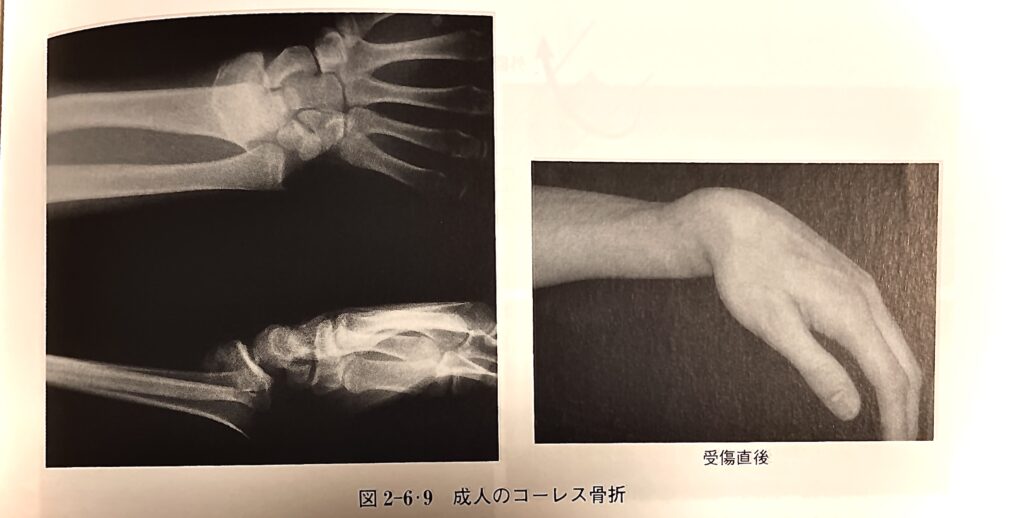
コーレス骨折
柔道整復学理論編 改訂第6版 より引用(一部改変)
多くは介達外力によって発生し、直達外力によって発生するものは稀である。手掌を衝いて転倒した際に、橈骨遠位端に受ける長軸圧と、手関節を含めて強度な伸展(背屈)力が強制され、橈骨遠位端部に掌側凸の屈曲力が働いて、手関節の1〜3㎝近位で骨折する。
骨折線は前額面では橈側近位から斜めに尺側遠位に走る。矢状面内では、手関節の1〜3㎝近位の掌側から、やや斜めに背側近位方向へ走る。
騎乗転移するとフォーク状変形を生じる。
このように、具体的にどのような骨折かの記載がありますね。
「手関節の1〜3㎝近位で骨折し、背側方向へ転位するものをコーレス骨折と言う」といったニュアンスで記載があります。
教科書の文面を見ると、転位の度合いはコーレス骨折の定義には含まれていないようですが、教科書の参考画像は、騎乗転位しているレントゲンが載っています。
もちろん、教科書は「コーレスさんはこのように発表しました」という内容を書いているわけではないので嘘はないのですが、実際にコーレスさんが発表したものとは違いますね。
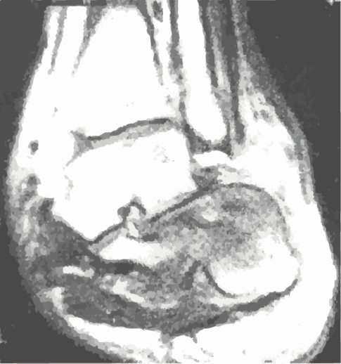
対して、コーレスさんが発表した骨折は、以下のようなものだと思います。
これを見ると、明らかに完全に騎乗転位している、もしくはそれに近い形で転位が強い橈骨遠位端骨折を想定しているものだと思います。
つまり、本当のコーレスさんが発表したコーレス骨折は、単純に背側方向へ転位する橈骨遠位端骨折のことをいうのではなく、外観上変形が明らかなものを言っているのだと判明しました。
コーレス骨折の定義は?
しかし、結論から言うと「コーレス骨折はこの骨折です」という定義はありません。
それは、そもそもコーレスさんが「これをコーレス骨折と呼びます」と言っていない上、その後にもコーレス骨折と定義する人は現れていないからです。
なんとなく骨折治療の界隈で「コーレスさんが言ってたあの骨折」というのがめんどくさくなり、いつのまにかコーレス骨折と呼ぶようになったのでしょう。
あくまでもコーレスさんは、「これまで得体の知れなかった手関節の怪我は実は骨折でした」という発表をしただけであって、橈骨遠位端骨折の型を発表したわけではなかったのですね。
まとめ
これはほねゆきの推測ですが、外観上の変形が明らかなものは、コーレスさんも骨折と判断していたのでしょうが、それほど転位が強くない橈骨遠位端骨折は骨折と言わずに、コーレスさんもそのまま捻挫などとして流していたのだと思います。
「変形しているけど中では何が起こっているのだろう?」→骨折だった
という発表ですから、変形があまりないものに関しては、適当に治療してもなんとなくなおるので、それを骨折と判明させる必要がきっとなかったので、コーレスさんは変形が著明なものに関して文章を書いたのでしょう。
今回は、時代背景や、その時に考えられていたものを知る事で、本当のコーレス骨折について迫ってみました。
みなさんは、どう思われましたか?
検査が潤沢に行える現代日本に、改めて感謝するべきなのかもしれません。
クーパー卿、ここで働かせてください!!!